Phone:
(701)814-6992
Physical address:
6296 Donnelly Plaza
Ratkeville, Bahamas.
Phone:
(701)814-6992
Physical address:
6296 Donnelly Plaza
Ratkeville, Bahamas.
Cone Beam Computed Tomography ( CBCT) is an advanced 3 D Imaging Modality that has high clinical applications for Dental and Maxillofacial Imaging.
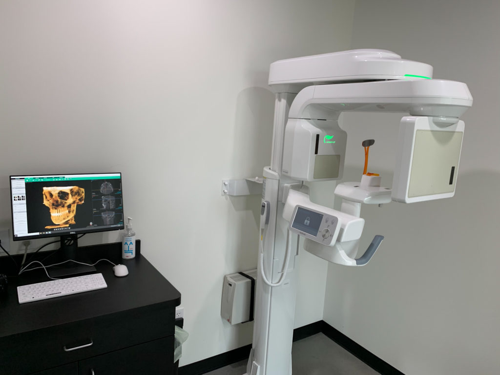
CBCT is highly accurate and is able to provide a 3 Dimensional volumetric data in Axial, Coronal and Sagittal Planes.
Radiation Exposure dose from CBCT is 10 times less than from Conventional CT scans during maxillofacial exposure.
Oralvision equipped with J Morita’s Veraview X 800 holds the capacity of procuring scans from 4×4 cm to 15X14 Cm. Offers a minute voxel size of just 80 micrometers providing higher resolution images.
This unit comes with 360 and 180 exposure modes, 360 degree mode can be used for greater acuracy and 180 degree mode can be used for lower X-Ray dose and a quicker exposure time.
A uniquely shaped Field of View ( FOV) with 10 cm encompasses the entire dental arch. Imaging of the entire arch can be executed with less X-Ray dose by excluding areas outside the region of interest.
The Veraview X 800’s largest field of view of 15 cm allows a scan of the entire jaw region.
Field of view refers to the anatomical area of the patient that will be exposed to radiation or the anatomical area that will be included in the data volume.
Oral vision equipped with J Morita’s Veraview X 800 provides the CBCT scans with FOV ranging from 4×4 cm to 15×14 cm.
Significant FOVs provided with Veraview X 800 are : 4×4 cm (in high resolution of 80 microns), 4×8 cm, 8×4 cm, 8×5 cm, 8×8 cm, 10×8 cm, ( in 125 microns) 15×5 cm and 15×14 cm ( in 320 microns).
Features
CBCT is highly accurate and can be used to provide the following procedures.
Here are the simple procedures for CBCT Imaging